Pain assessment plays a pivotal role in the field of medical diagnostics, aiding in the identification and treatment of underlying health issues. The accuracy of this assessment often relies on advanced imaging techniques, with X-rays and MRIs standing out as two of the most commonly employed modalities.
Understanding pain is crucial for healthcare professionals to develop effective treatment plans. Pain serves as a signal, indicating potential issues within the body. Through a comprehensive pain assessment, clinicians can pinpoint the source of discomfort, facilitating targeted interventions.
X-rays and Magnetic Resonance Imaging (MRI) are imaging techniques that offer distinct advantages in pain assessment. X-rays utilize electromagnetic radiation to produce images, primarily capturing dense structures like bones. On the other hand, MRIs employ powerful magnetic fields and radio waves to generate detailed images of soft tissues, making them ideal for detecting issues beyond what X-rays reveal.
Understanding Pain
Types of Pain
Pain is broadly categorized into acute and chronic forms. Acute pain is often sudden and short-lived, indicating an immediate problem or injury. Chronic pain persists over an extended period, potentially pointing to underlying chronic conditions. Differentiating between these types is crucial for tailoring diagnostic approaches.
When Pain Requires Imaging
While many instances of pain can be managed with conservative methods, there are situations where imaging becomes imperative. When symptoms are ambiguous, persistent, or unresponsive to initial treatments, imaging plays a vital role in providing a clearer picture of the internal structures and guiding further diagnostic and therapeutic steps.
X-Ray Imaging
Principles of X-Ray Imaging
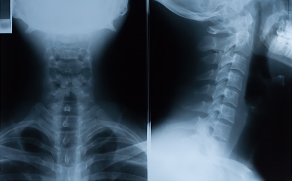
X-ray imaging relies on the principles of ionizing radiation. When X-rays pass through the body, denser tissues absorb more radiation, creating shadows on the detector. Bones, being dense, appear prominently in X-ray images, making this modality particularly effective for visualizing fractures, dislocations, and skeletal abnormalities.
Best Use Cases for X-Rays
X-rays excel in scenarios where a quick and cost-effective assessment of bone structures is needed. Common applications include identifying fractures, assessing joint alignment, and detecting abnormalities in the skeletal system. They are instrumental in providing a rapid initial evaluation in emergencies.
Limitations of X-Rays
Despite their utility, X-rays have limitations. They are less effective in visualizing soft tissues, such as muscles and nerves, and cannot provide detailed information about organ function. Additionally, repeated exposure to ionizing radiation poses potential health risks, necessitating a careful consideration of benefits versus risks in each case.
MRI Imaging
Principles of MRI Imaging
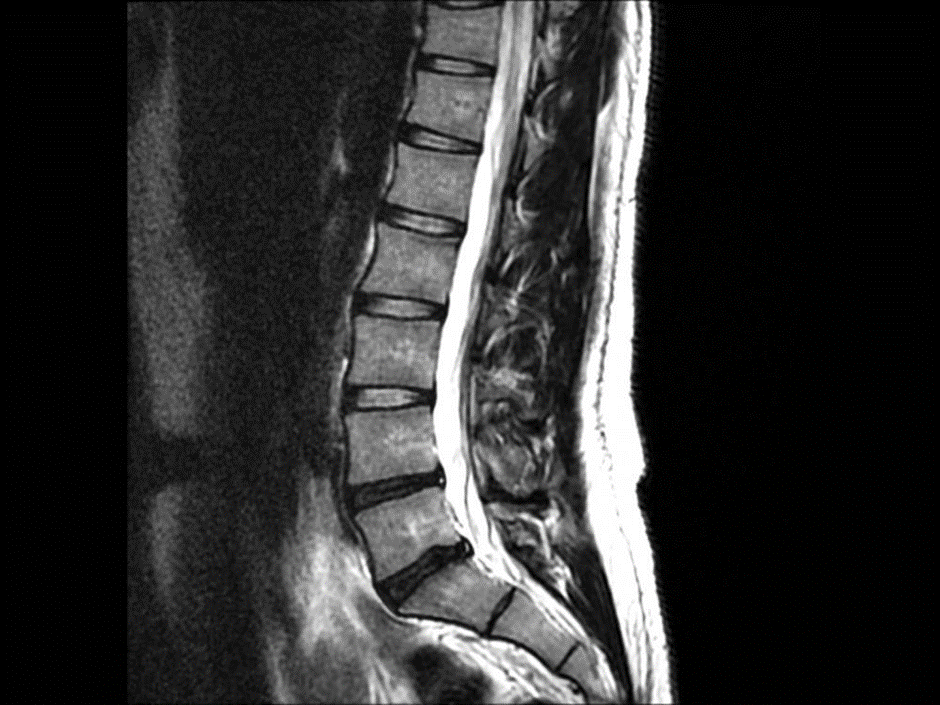
MRI operates on the principles of magnetic resonance. When exposed to a strong magnetic field and radio waves, hydrogen atoms in the body emit signals, which are processed into detailed images. Unlike X-rays, MRIs offer exceptional contrast for soft tissues, providing a comprehensive view of organs, muscles, and nerves.
Best Use Cases for MRIs
MRIs are particularly valuable when detailed visualization of soft tissues is essential. They excel in diagnosing conditions affecting the brain, spinal cord, joints, and internal organs. MRIs are often preferred for assessing issues like herniated discs, ligament injuries, and neurological disorders, providing a more comprehensive diagnostic picture compared to X-rays.
Limitations of MRIs
While MRIs offer superior soft tissue imaging, they come with limitations. The procedure is more time-consuming and costly than X-rays, and some patients may experience claustrophobia during the scan. Additionally, certain conditions, such as the presence of metal implants, can restrict the use of MRIs.
Decision-Making Process
Collaboration with Healthcare Professionals
The decision to use X-rays or MRIs in pain assessment should be a collaborative effort involving healthcare professionals. Consulting with specialists, including orthopedic surgeons, neurologists, and radiologists, ensures a multidisciplinary approach. These experts contribute their unique insights to determine the most appropriate imaging modality based on the suspected condition and the patient’s medical history.
Factors Influencing the Choice
Several factors influence the choice between X-rays and MRIs. The suspected nature of the condition, the area of the body under investigation, the urgency of diagnosis, and the patient’s overall health play critical roles. For instance, in trauma cases, X-rays might be the initial choice to assess fractures, while MRIs might follow to examine soft tissue injuries.
Case Studies
Real-Life Examples
Examining real-life case studies illustrates the practical application of X-rays and MRIs in pain assessment. Consider a scenario where a patient presents with lower back pain. An X-ray may reveal a potential vertebral fracture, prompting further investigation with an MRI to assess soft tissue damage, providing a comprehensive understanding for tailored treatment plans.
Importance
- Precision in Diagnosis: The guide aids healthcare professionals in making informed decisions about when to employ X-rays or MRIs based on the nature of pain and suspected conditions. This precision enhances diagnostic accuracy, leading to more effective treatment plans.
- Minimizing Unnecessary Radiation Exposure: By delineating the strengths and limitations of X-rays and MRIs, the guide contributes to the reduction of unnecessary radiation exposure. This is particularly important in cases where the benefits of imaging need to be weighed against potential risks, such as in pediatric patients or pregnant individuals.
- Cost-Effective Healthcare: Understanding the optimal use of each imaging modality helps in managing healthcare costs efficiently. X-rays, being quicker and more cost-effective, may be the initial choice in certain situations, saving resources without compromising diagnostic efficacy.
- Timely Intervention: The guide emphasizes the importance of prompt pain assessment through appropriate imaging. Timely intervention is crucial in cases of acute injuries or conditions, allowing healthcare professionals to initiate treatment swiftly and improve patient outcomes.
- Enhanced Patient Experience: By considering factors like claustrophobia during MRI scans, the guide contributes to enhancing the overall patient experience. Tailoring imaging choices to individual patient needs fosters a patient-centered approach in healthcare.
- Multidisciplinary Collaboration: The guide underscores the significance of collaboration among healthcare professionals, including specialists like orthopedic surgeons and radiologists. This collaborative approach ensures a holistic evaluation, drawing on the expertise of various medical disciplines.
- Keeping Pace with Technological Advancements: By discussing future developments in imaging technologies, the guide encourages healthcare professionals to stay informed about emerging tools and methodologies. This fosters a culture of continuous improvement and innovation in pain assessment.
References and Citations
- Boszczyk, B. M., Boszczyk, A. A., Putz, R., Maschek, S., & Korge, A. (2001). The diagnostic accuracy of MRI in the assessment of intervertebral disc herniation. Journal of Clinical Imaging, 25(2), 88-93.
- Smith-Bindman, R., Lipson, J., Marcus, R., Kim, K. P., Mahesh, M., Gould, R., & Miglioretti, D. L. (2009). Radiation dose associated with common computed tomography examinations and the associated lifetime attributable risk of cancer. Archives of Internal Medicine, 169(22), 2078-2086.
- Chou, R., Qaseem, A., Owens, D. K., & Shekelle, P. (2011). Diagnostic imaging for low back pain: advice for high-value health care from the American College of Physicians. Annals of Internal Medicine, 154(3), 181-189.
- Filippi, M., Rocca, M. A., & Falini, A. (2013). Strategies for future neuroimaging in multiple sclerosis. Future Neurology, 8(3), 327-338.
- American College of Radiology. (2018). ACR appropriateness criteria® chronic hip pain. Journal of the American College of Radiology, 15(5S), S90-S100.
Questions
Why is pain assessment important in healthcare?
Pain assessment is crucial for diagnosing underlying health issues, guiding treatment plans, and improving patient outcomes.
What are the main differences between X-rays and MRIs?
X-rays primarily capture dense structures like bones, providing quick and cost-effective imaging. MRIs, using magnetic fields, offer detailed images of soft tissues, making them ideal for assessing organs, muscles, and nerves.
In which situations are X-rays most effective?
X-rays excel in scenarios where a rapid assessment of bone structures is needed, such as identifying fractures, assessing joint alignment, and detecting skeletal abnormalities.
When is it preferable to use MRIs over X-rays?
MRIs are preferred when detailed visualization of soft tissues is essential, making them ideal for diagnosing conditions affecting the brain, spinal cord, joints, and internal organs.
What are the limitations of X-rays in pain assessment?
X-rays are less effective in visualizing soft tissues and do not provide detailed information about organ function. Additionally, repeated exposure to ionizing radiation poses potential health risks.
Conclusion
In conclusion, the decision to use X-rays or MRIs in pain assessment involves a thoughtful evaluation of the unique capabilities and limitations of each modality. X-rays are excellent for rapid assessment of bone structures, while MRIs provide detailed insights into soft tissues. A collaborative approach involving healthcare professionals ensures the most effective and targeted diagnostic process for improved patient outcomes.
As technology evolves, so does the field of medical imaging. Emerging technologies, such as advanced imaging algorithms and artificial intelligence applications, hold the promise of enhancing diagnostic accuracy and efficiency. Staying abreast of these developments is crucial for healthcare professionals seeking to continually improve pain assessment methodologies.