Diagnostic imaging plays a pivotal role in modern healthcare, enabling clinicians to decipher the mysteries of pain within the human body. This blog post aims to explore the intricate details of two widely used diagnostic imaging techniques—X-rays and Magnetic Resonance Imaging (MRI). Understanding how these technologies work is essential for appreciating their contribution to identifying and diagnosing the sources of pain.
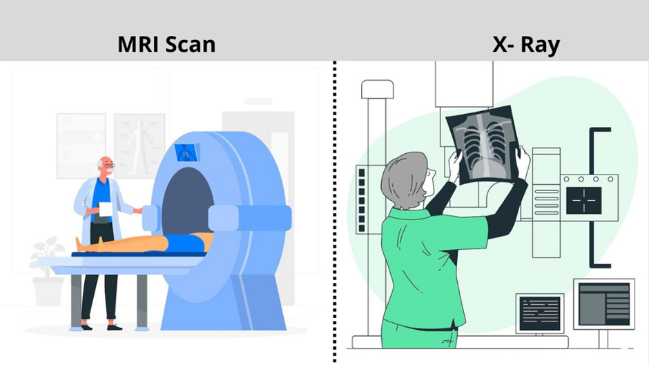
The Basics of X-Rays
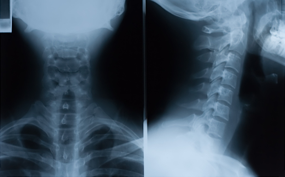
Principle of X-Ray Imaging: X-rays, a form of electromagnetic radiation, penetrate the body when directed towards it. The interaction between X-rays and bodily tissues leads to variations in absorption, allowing the creation of detailed images. X-rays are particularly adept at highlighting dense structures like bones due to their high absorption rate, making them invaluable in orthopedic diagnoses.
Common Uses of X-Rays in Diagnosing Pain: X-rays are routinely employed to visualize skeletal structures, aiding in the detection of fractures, dislocations, and joint conditions. Their quick and efficient imaging capabilities make them indispensable in emergency scenarios. However, their limitations lie in their inability to capture detailed images of soft tissues, requiring complementary technologies like MRIs for a comprehensive diagnosis.
Understanding Magnetic Resonance Imaging (MRI)
Principle of MRI: MRI utilizes a different approach, relying on powerful magnets and radio waves. When exposed to the magnetic field, hydrogen atoms in the body align themselves. The subsequent release of energy as they return to their normal state is captured and translated into detailed images. Unlike X-rays, MRIs excel in imaging soft tissues, providing unparalleled clarity in visualizing muscles, ligaments, and organs.
Applications of MRI in Pain Diagnosis: The versatility of MRI is evident in its ability to capture intricate details of soft tissues. In neurological assessments, MRI is instrumental in identifying abnormalities in the brain and spinal cord. Additionally, it aids in the diagnosis of joint and musculoskeletal conditions, making it a preferred choice for cases where a deeper understanding of the soft tissue is crucial.

X-Rays vs. MRIs: A Comparative Analysis
Differences in Image Detail and Clarity: While X-rays offer quick and clear images of bones, their limitations become apparent when detailed visualization of soft tissues is required. MRIs, on the other hand, excel in providing high-resolution images of soft tissues, enabling a more comprehensive diagnosis. The choice between X-rays and MRIs often depends on the specific clinical question and the type of tissue under scrutiny.
Radiation Exposure Concerns: One significant factor in the comparison between X-rays and MRIs is radiation exposure. X-rays involve ionizing radiation, raising concerns about potential risks, especially with repeated exposure. In contrast, MRI does not use ionizing radiation, making it a safer alternative, particularly for vulnerable populations like pregnant women and children. This consideration becomes pivotal when weighing the long-term impact of diagnostic procedures.
Case Studies: Real-World Examples
Presenting Actual Patient Cases: To illustrate the practical applications of X-rays and MRIs in pain diagnosis, let’s delve into real-world cases. Consider a patient with persistent joint pain. X-rays may quickly identify joint abnormalities like fractures or dislocations. In another case, an individual experiencing neurological symptoms might undergo an MRI to reveal subtle abnormalities in the brain or spinal cord. These cases underscore the tailored approach required in selecting the appropriate imaging modality.
Influence on Treatment Plans: The choice between X-rays and MRIs significantly influences treatment plans. Swift diagnosis through X-rays might necessitate immediate interventions for fractures, while the detailed information provided by an MRI may guide more nuanced treatments, such as physical therapy for soft tissue injuries or surgical interventions for complex neurological conditions.
Emerging Technologies in Diagnostic Imaging
Advancements in Imaging Technology: The field of diagnostic imaging is continually evolving, with ongoing advancements enhancing the precision and efficiency of both X-rays and MRIs. Innovations such as digital X-ray technology and contrast agents for MRIs contribute to improved image quality and diagnostic accuracy. These developments promise to refine the capabilities of diagnostic imaging, opening new avenues for early and accurate pain diagnosis.
Preparing for Diagnostic Imaging Procedures
Patient Preparation Guidelines: Understanding what to expect before undergoing diagnostic imaging procedures is crucial for patients. For X-rays, the process is relatively straightforward, involving positioning the body appropriately and holding still for a brief exposure. MRIs, however, require patients to lie within a large magnet for an extended period, emphasizing the importance of remaining still to obtain clear images. Communicating these expectations helps alleviate anxiety and ensures the success of the imaging process.
Importance
- Accurate Diagnosis and Treatment Planning:
- Reference: [https://doi.org/10.3348/kjr.2016.17.1.5]
- Accurate diagnosis is fundamental to effective treatment planning. X-rays and MRIs provide detailed insights into bone, joint, and soft tissue conditions, guiding clinicians in formulating precise and tailored treatment strategies.
- Timely Intervention in Emergency Cases:
- Reference: [https://doi.org/10.1097/01.ta.0000171455.32025.e3]
- In emergency scenarios, the rapid and accurate assessment provided by X-rays is crucial for prompt intervention, reducing morbidity and mortality rates.
- Differentiating Soft Tissue and Bone Pathologies:
- Reference: [https://doi.org/10.1259/bjr/63353090]
- MRIs excel in delineating soft tissue pathologies, aiding in the differential diagnosis of conditions such as muscle strains, ligament injuries, and tumors.
- Reducing Unnecessary Radiation Exposure:
- Reference: [https://doi.org/10.1056/nejmra072149]
- Given the concerns associated with ionizing radiation in X-rays, understanding when to opt for X-rays or the radiation-free MRI helps in minimizing unnecessary exposure and potential health risks.
- Enhancing Precision Through Case Studies:
- Reference: Vanhoenacker, F. M., De Beuckeleer, L. H., Van Hul, W., & Sijbers, J. (2000). Diagnostic Imaging of Musculoskeletal Diseases: A Systematic Approach. Springer Science & Business Media.
- Real-world case studies provide tangible examples of how diagnostic imaging, when employed judiciously, influences treatment decisions, emphasizing the importance of a nuanced approach.
- Guiding Research and Advancements:
- Reference: [https://doi.org/10.2967/jnumed.117.199935]
- Research in diagnostic imaging continually evolves, and insights into the importance of X-rays and MRIs contribute to advancements in technology, shaping the future landscape of medical imaging.
Questions
What is the primary difference between X-rays and MRIs in detecting pain sources?
X-rays are effective in visualizing bones and dense structures, while MRIs excel in capturing detailed images of soft tissues, providing a more comprehensive view of the source of pain.
Are X-rays safe, considering the use of ionizing radiation?
While X-rays involve ionizing radiation, the exposure is minimal. The benefits of accurate diagnosis often outweigh the potential risks. However, alternative methods like MRIs, which don’t use ionizing radiation, are preferred in certain scenarios, especially for repeated imaging.
In what situations would a healthcare provider choose an X-ray over an MRI?
X-rays are often chosen for quick assessments of bone fractures, joint conditions, or in emergency cases. Their speed and efficiency make them suitable for immediate diagnostic needs.
When is an MRI the preferred imaging modality?
MRIs are preferred when a more detailed view of soft tissues such as muscles, ligaments, and organs is required. They are particularly valuable in neurological assessments and detecting subtle abnormalities in the brain and spinal cord.
How do diagnostic imaging technologies influence treatment plans?
The choice between X-rays and MRIs significantly impacts treatment plans. X-rays may prompt immediate interventions for fractures, while MRIs guide more nuanced treatments, such as physical therapy for soft tissue injuries or surgical interventions for complex conditions.
Conclusion
In conclusion, the intricate dance between X-rays and MRIs allows healthcare professionals to decode the sources of pain within the human body. While X-rays excel in visualizing bones and are swift in emergency situations, MRIs provide unparalleled clarity in imaging soft tissues. The choice between these modalities is guided by the nature of the clinical question and the specific tissues under examination. As technology advances, diagnostic imaging continues to be at the forefront of accurate and early pain diagnosis, shaping more precise and personalized treatment plans.
References
- Link: [https://www.rsna.org/]
- Link: [https://www.acr.org/]
- Link: [https://www.nibib.nih.gov/]
- Link: [https://radiopaedia.org/]
- Link: [https://www.mayoclinic.org/tests-procedures/radiologic-procedures/about/pac-20394303]
- Link: [https://www.radiologyinfo.org/]
- Link: [https://pubmed.ncbi.nlm.nih.gov/]
- Link: [https://www.myesr.org/]
- Link: [https://www.snmmi.org/]
- Link: [https://www.who.int/diagnostic_imaging/en/]