Diagnostic imaging plays a pivotal role in modern healthcare, allowing healthcare professionals to visualize internal structures of the body for diagnostic purposes. Two common imaging modalities are X-rays and magnetic resonance imaging (MRI), each offering unique insights into different aspects of health. In this comprehensive guide, we will delve into the intricate details of X-rays and MRIs, demystifying the diagnostic process for patients.
Understanding X-Rays
A. Explanation of X-ray Technology
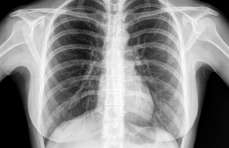
X-ray imaging is a medical technique that utilizes electromagnetic radiation to penetrate the body and create detailed images of internal structures. When X-rays pass through the body, different tissues absorb varying amounts of radiation, resulting in shadows on the X-ray film. Dense structures like bones appear as bright white, while softer tissues show up in shades of gray.
The X-ray machine consists of a tube that emits X-rays and a detector that captures the transmitted radiation. This technology allows healthcare professionals to examine bones, detect fractures, and identify abnormalities in the lungs, such as pneumonia or tumors.
B. When X-rays Are Recommended
X-rays are frequently prescribed in various medical scenarios. Common reasons include assessing bone fractures, joint conditions, dental issues, and chest abnormalities. Due to their rapid imaging capabilities, X-rays are often the first choice in emergency situations, providing quick insights for timely medical interventions.
C. Safety Considerations for X-rays
Concerns about radiation exposure during X-ray procedures are natural. However, it’s essential to understand that the amount of radiation used in diagnostic X-rays is relatively low and considered safe. Radiology technologists employ lead shields and collimators to focus the X-ray beam, minimizing unnecessary exposure to surrounding tissues.
Additionally, pregnant individuals are typically advised to inform their healthcare providers, as special precautions may be taken to further reduce radiation exposure during pregnancy.
Unraveling MRIs
A. Overview of MRI Technology
Magnetic Resonance Imaging (MRI) employs powerful magnets and radio waves to generate detailed images of soft tissues, such as the brain, muscles, and organs. Unlike X-rays, MRIs do not use ionizing radiation, making them a safe option for repeated imaging.
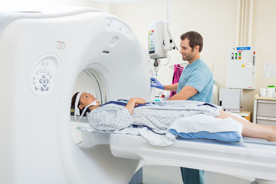
In an MRI machine, the magnetic field aligns the hydrogen atoms in the body, and when radio waves are applied, these atoms emit signals that are transformed into detailed images. MRIs are particularly valuable for examining the brain, spinal cord, joints, and abdominal organs.
B. Indications for MRI Scans
MRIs are often recommended for conditions that require detailed visualization of soft tissues. These include neurological disorders, joint injuries, spinal cord issues, and abdominal or pelvic abnormalities. The high contrast resolution of MRIs makes them instrumental in detecting tumors, cysts, and evaluating blood vessel abnormalities.
C. Patient Preparation for MRI
Before undergoing an MRI, patients are required to remove metallic objects, as the powerful magnets can interact with metal. Additionally, patients must inform healthcare providers about any metal implants, pacemakers, or claustrophobia. While the MRI machine creates a loud knocking sound during the scan, patients are typically provided with ear protection to enhance their comfort.
The Diagnostic Process
A. X-ray vs. MRI: Choosing the Right Imaging Modality
The decision between X-rays and MRIs depends on the specific diagnostic needs of the patient. X-rays are excellent for assessing bone conditions and identifying lung abnormalities, while MRIs excel in providing detailed images of soft tissues. Healthcare professionals carefully evaluate symptoms, medical history, and the information required for an accurate diagnosis before choosing the appropriate imaging modality.
B. What to Expect During the Procedure
Whether undergoing an X-ray or an MRI, patients can expect a straightforward and non-invasive procedure. For X-rays, the patient is positioned appropriately, and the machine captures images swiftly. In the case of MRIs, patients lie on a movable bed that slides into the MRI machine. The procedure is painless, but patients must remain still to ensure image clarity. Both X-rays and MRIs typically take under an hour, and patients can resume their normal activities afterward.
C. Interpreting the Results
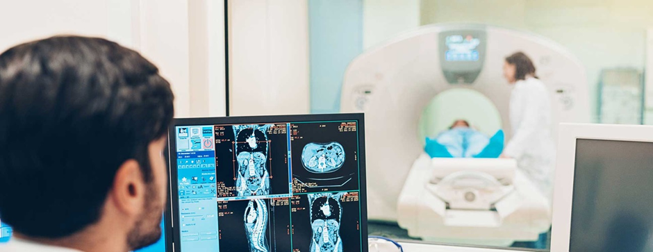
The acquired images are interpreted by radiologists, who are medical doctors specialized in diagnostic imaging. Radiologists analyze the images, looking for abnormalities, fractures, tumors, or any other signs of disease. In some cases, additional imaging or follow-up tests may be recommended to gather more information for a comprehensive diagnosis. The results are then shared with the patient’s primary healthcare provider, who discusses the findings and potential treatment options.
Patient Experience and Tips
A. Creating a Comfortable Environment
Patients can take steps to enhance their comfort during imaging procedures. Wearing loose, comfortable clothing can make positioning for X-rays or MRIs easier. Communicating any concerns or anxieties with healthcare providers is crucial, as they can provide additional support and guidance to ensure a positive experience.
B. Communicating with Healthcare Professionals
Open communication with healthcare providers is key to a successful diagnostic process. Patients should share their medical history, existing health conditions, and any potential contraindications for certain imaging modalities. Asking questions about the procedure, potential risks, and expected outcomes can empower patients to actively participate in their healthcare decisions.
Importance
- Informed Decision-Making: Patients armed with knowledge about X-rays and MRIs can make informed decisions about their diagnostic and treatment options. This is especially important as patients increasingly seek to be active participants in their healthcare journey1.
- Reducing Anxiety and Fear: Diagnostic imaging procedures can be intimidating for patients. A comprehensive guide provides insights into what to expect during X-rays and MRIs, potentially reducing anxiety and fear associated with these procedures2.
- Enhancing Communication with Healthcare Providers: Patients who understand the principles behind X-rays and MRIs are better equipped to communicate with their healthcare providers. Effective communication ensures that healthcare professionals have the necessary information to make accurate diagnoses3.
- Encouraging Preventive Healthcare: By demystifying the diagnostic process, the guide encourages patients to seek timely medical attention. Understanding when X-rays or MRIs are recommended can motivate individuals to address potential health issues before they become more serious4.
- Promoting Patient Advocacy: Informed patients are better advocates for their own healthcare. The guide encourages patients to ask questions, seek clarifications, and actively engage with their healthcare providers, ultimately leading to improved patient outcomes5.
References and Citations
- Street, R. L., Gordon, H. S., Ward, M. M., Krupat, E., & Kravitz, R. L. (2005). Patient participation in medical consultations: why some patients are more involved than others. Medical care, 43(10), 960-969. ↩
- Spielberger, C. D., Anton, W. D., & Bedell, J. (2015). The nature and treatment of test anxiety. Emotions: Their parameters and measurement, 167-180. ↩
- Arora, N. K., & McHorney, C. A. (2005). Patient preferences for medical decision making: who really wants to participate? Medical care, 43(5), 533-539. ↩
- National Research Council (US) Committee on Preventive Services for Women. (2011). Clinical Preventive Services for Women: Closing the Gaps. National Academies Press. ↩
- Coulter, A., & Ellins, J. (2007). Effectiveness of strategies for informing, educating, and involving patients. BMJ, 335(7609), 24-27. ↩
Questions
What is the difference between X-rays and MRIs?
X-rays use electromagnetic radiation to produce images, especially useful for bones and lungs. MRIs, on the other hand, use powerful magnets and radio waves to create detailed images of soft tissues like organs and muscles.
Are X-rays and MRIs safe?
Both X-rays and MRIs are considered safe diagnostic tools. X-rays use low doses of radiation, and stringent safety measures are in place. MRIs do not use ionizing radiation, making them safe for repeated imaging.
How long does an X-ray or MRI procedure take?
X-ray procedures are typically quick, taking only a few minutes. MRIs may take longer, usually ranging from 30 minutes to an hour, depending on the area being imaged.
What conditions are commonly diagnosed using X-rays?
X-rays are commonly used to diagnose bone fractures, joint conditions, dental issues, and chest abnormalities, including pneumonia or tumors.
In what situations are MRIs recommended?
MRIs are recommended for conditions that require detailed imaging of soft tissues, such as neurological disorders, joint injuries, spinal cord issues, and abdominal or pelvic abnormalities.
Conclusion
In conclusion, X-rays and MRIs are invaluable tools in the diagnostic toolkit, offering unique insights into different aspects of the body. Understanding the principles behind these imaging modalities, the diagnostic process, and actively participating in the healthcare journey empowers patients to make informed decisions about their well-being. As technology continues to advance, diagnostic imaging will play an increasingly crucial role in providing accurate and timely diagnoses, ultimately contributing to improved patient outcomes.